
What composes the hip joint?
Health information
Components of the Hip Joint
What is the Hip Joint?
The hip joint is a crucial joint that serves as the center for all movements, including standing, walking, and running. This article will provide a detailed explanation of the structure of the hip joint.
1. The Skeletal Structure of the Hip Joint
The hip joint is classified as a ball-and-socket joint that connects the pelvis and the leg. It is the second most mobile joint in the human body, after the shoulder joint. It is a flexible joint capable of planar movements such as flexion and extension, as well as three-dimensional movements like circumduction.
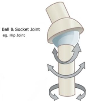
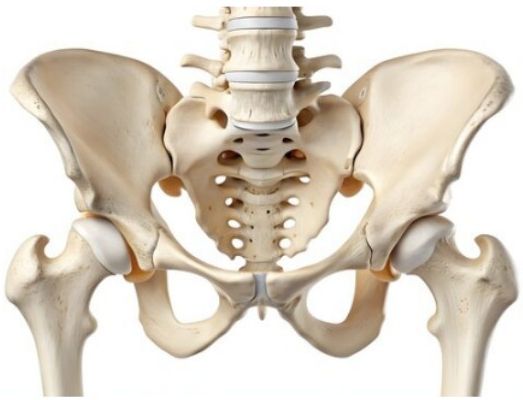
This joint is formed by the articulation of the femoral head and the acetabulum of the pelvic bone. As it must support the body's weight, it is stronger, more stable, and reinforced by numerous muscles and ligaments compared to the shoulder joint, which is also a ball-and-socket joint.
2. The Muscular Structure of the Hip Joint
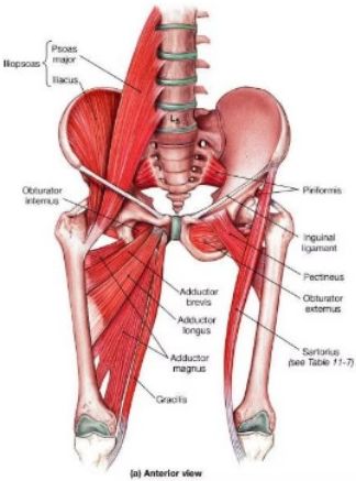
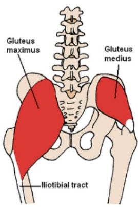
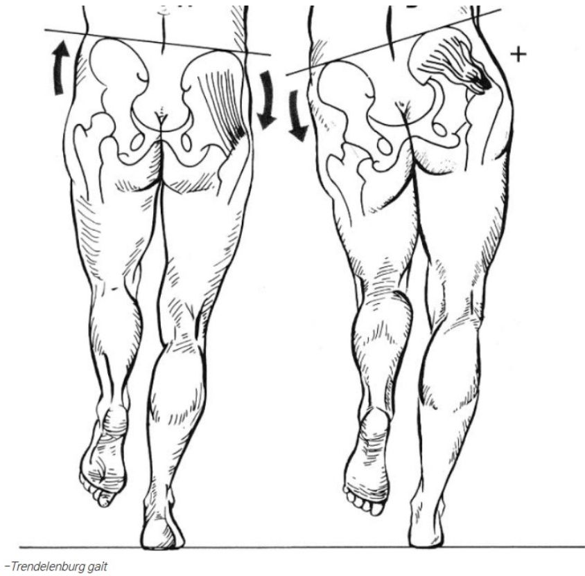
As previously mentioned, the hip joint is reinforced by various muscles and ligaments. While the provided images do not encompass all the muscles, they effectively illustrate the primary ones. Notably, among the posterior muscles, the gluteus medius is arguably the most important muscle of the hip joint.
This muscle is the site for intramuscular injections in the buttocks. The gluteus medius plays a vital role in walking by abducting and internally rotating the hip joint. Therefore, weakness or damage to this muscle can result in a distinctive waddling motion known as the Trendelenburg gait.

3. The Nerves of the Hip Joint
The hip joint, also referred to as the pelvis, is a critical biological location that houses various spinal nerves and organs. It is the most frequently injured area in falling accidents and has the poorest prognosis following such incidents.
Lumbar Plexus
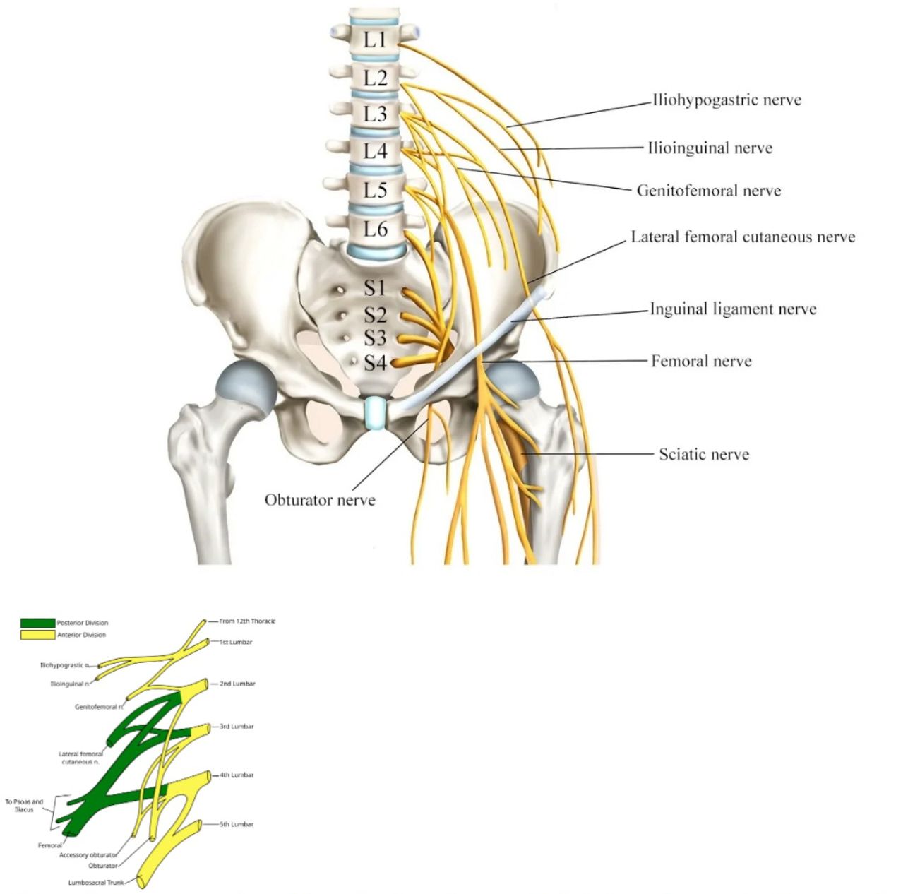
- Iliohypogastric nerve
- Ilioinguinal nerve
- Genitofemoral nerve
- Lateral femoral cutaneous nerve
- Inguinal ligament nerve
- Femoral nerve
- Sciatic nerve
- Obturator nerve
In fact, research indicates a very high mortality rate within one year for patients who have undergone Total Hip Replacement (THR) surgery after a hip fracture. As can be seen in the illustration, complex nerve bundles extend from the spinal cord. Consequently, any injury to the hip joint inevitably leads to neurological and musculoskeletal problems.
4. The Ligaments of the Hip Joint
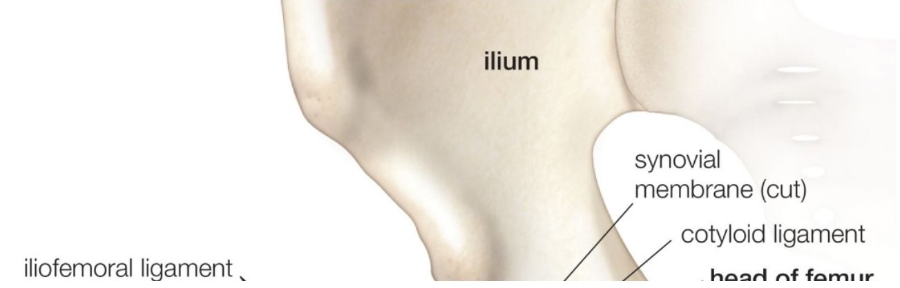
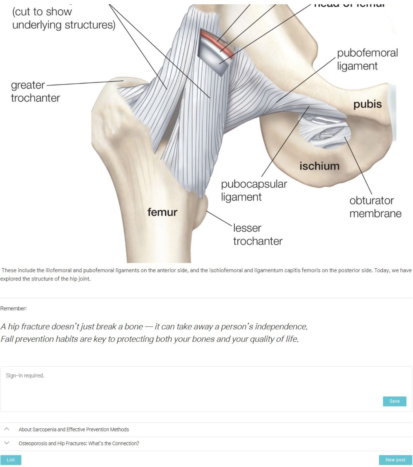
It was stated earlier that the hip joint is formed by the articulation of the femoral head and the acetabulum. While the primary advantage of a ball-and-socket joint is its mobility, this freedom can lead to instability. To counteract this, the human body reinforces the hip joint with several ligaments.
💪 Remember
A hip fracture doesn't just break a bone — it can take away a person's independence.
Fall prevention habits are key to protecting both your bones and your quality of life.